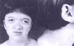
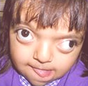
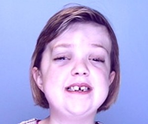
Crouzon syndrome is a genetic disorder. It is one of many birth defects that results in abnormal fusion between bones in the skull and face. Normally, as an infant’s brain grows, open sutures between the bones, allow the skull to develop normally. When sutures fuse too early, the skull grows in the direction of the remaining open sutures. In Crouzon syndrome, bones in the skull and face fuse too early, resulting in an abnormally shaped head, face, and teeth.
It is a genetic autosomal dominant disorder, caused by mutations of the FGFR2 (fibroblast growth factor receptor), or by less common FGFR3 genes. A mutation in these genes may cause bones in the skull to fuse too early.
The craniofacial team has treated 31 children in Seattle with Crouzon syndrome in past 5 years. Every year they receive two new children for the treatment of Crouzon syndrome, and perform about 13 surgeries in each year.
Symptoms of Crouzon syndrome
Some of the signs and symptoms of Crouzon syndrome are listed below:
- Lack of alignment between the two eyes which may affect the vision
- Protrusion of the eyeballs from the eye socket due to shallow eye sockets
- Increase in distance between the two eyes
- Low- set ears, associated with partial or complete loss of hearing. Some severe cases may present with Meniere’s disease.
- Beak like appearance of the nose
- Inadequate growth of mid part of the face and smaller upper jaw
- Tall narrow palate
- Protrusion of chin
- Difficulty in breathing
- Abnormal skull size and facial features
- Dark, pigmented patches on neck, eyelids and around the mouth
- No defects in hands and legs
- Shorter femur and humerus bones
- Partial syndactyly
Types of Crouzon syndrome fusions
Crouzon Syndrome is caused due to the mutation of fibroblast growth factor receptor II and III, located on chromosome 10. The condition is characterized by premature fusion of the bones of the skull and face, which limits the ability of the affected bone to grow and expand.
The different patterns of fusion are:
- There is fusion of the coronal structure which leads to the formation of a flattened head or brachycephaly. The early fusion of the frontal bones to the two parietal bones also leads to a shortening of the skull diameter when measures from front to the back.
- There is fusion of the metopic suture leading to the development of a triangle-shaped forehead. It leads to fusion of the two frontal lobes which restricts the crossways growth, which eventually to a parallel development of the skull, giving it a triangular shape
- There is fusion of the sagittal suture which results in the development of a narrow and long head. It is caused due to early merging of the two parietal bones leading to an appearance similar to that of an inverted boat
- There is fusion of the coronal and lambdoid suture leading to an asymmetrical deformation of the skull. The merging of the skull bones causes the skull bones on one side to become increasingly flattened.
- There is fusion of all the sutures leading to the development of Kellebalttscheaedel
- There is fusion of the coronal and lamdboidal suture leading to the development of a high head. This is generally caused when the parietal and occipital bones get merged
Diagnosis of Crouzon syndrome
The following ways are used to diagnose Crouzon syndrome:
- The physical symptoms shown above and the characteristic facial features
- Display of defect in CT scan
- X-ray
- Test of sample cells, genetic tests and
- A study of the family’s medical history helps the doctor to diagnose the syndrome.
Causes of Crouzon syndrome
- A genetic change or mutation in fibroblast growth factor receptor genes-FGFR2 on chromosome 10 or FGFR3 on chromosome 4, causes the Crouzon syndrome. This syndrome can be inherited from any one of the parents, with a fifty percent chance of passing the mutation.
- Crouzon syndrome is different from other craniosynostosis syndromes because it does not cause abnormalities in hands and legs. But the cervical spine defects are common.
- Crouzon syndrome is a rare disorder, which affects about 1.6 per 100,000 persons, and about 4.5% among the subjects of craniosynostosis disorder.
Crouzon syndrome treatment
Surgical intervention is followed to correct the abnormalities associated with Crouzon syndrome
The major deformities of Crouzon Syndrome treatment plan involve correction via plastic surgery, which is also known as craniofacial surgery. The surgery is performed by a plastic surgeon with the support of neurosurgeons.
The types of surgery required are:
- To correct the mid face deficiency, plastic surgeons would move the lower orbit and the maxillary bone, forward
- Shaping the abnormalities of eye sockets and shallow orbits, and cranial surgery, to address the hydrocephalus condition, Chiari malfunction, abnormal ear canal, speech and breath disorders, sleep apnea, etc.
- Realigning of the eyes by reshaping the orbital cavity and moving the orbits forward
- Upper jaw surgery performed by a plastic surgeon, and for correction of other deformities within the oral cavity, such as high arched palate, bilateral cross bite, hypodontia or crowding of teeth.
Since the condition tends to affect more than one suture, an open surgery is preferred. Subsequent re-correction surgery is also a part of the Crouzon Syndrome treatment regimen.
Crouzon syndrome pictures


